In a 2016 U.K. survey, fewer than one-third of women were able to correctly name and identify six parts of their own reproductive system, according to the book “Pain and Prejudice: How the Medical System Ignores Women — And What We Can Do About It,” by Gabrielle Jackson, published in 2021. If you’re wondering if you know how to identify your body parts, you find yourself in good company: Forty-four percent of the women surveyed could not correctly identify the vagina, and 60% did not correctly label the vulva.
This is problematic when we as women try to discuss issues with our gynecologists and don’t have the proper terms, descriptions or understanding of our bodies to articulate what we are experiencing and where.
Because women’s health and words that refer to parts of women’s bodies are often considered taboo, not having the knowledge to talk about our own bodies is not something to be ashamed of; instead, it’s an opportunity to ask questions in order to learn to correctly name the parts of our bodies, so we can experience knowledgeably in them and speak confidently about them.
And if you’re a man, it’s important for you to know the correct words for women’s body parts, too. We’re happy you’re here.
Here are six parts of the female reproductive system, along with descriptions of each part’s function:
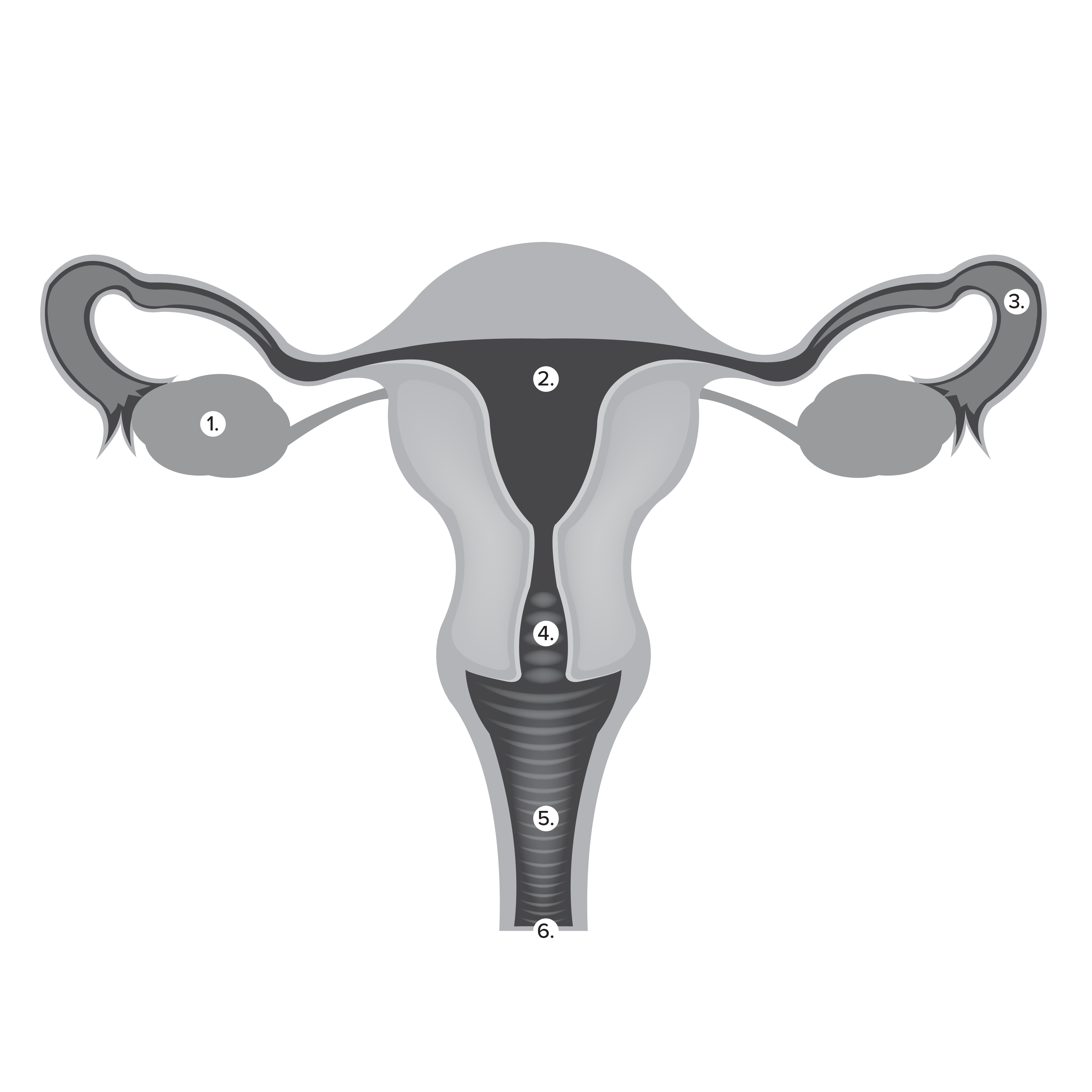
1. Ovaries: The primary female reproductive organs, these gonads about the size and shape of almonds release a mature egg each month from alternating sides, an event called ovulation. They also produce the main female sex hormones estrogen and progesterone, as well as a small amount of testosterone.
2. Uterus (Womb): A hollow organ about the size and shape of an upside-down pear inside of the pelvic cavity between the bladder and rectum. The place where a fetus grows before birth, the uterine lining is called the endometrium; when a woman isn’t pregnant, this lining sheds approximately once a month, known as a period. When a woman is pregnant, the fetus attaches to and is nourished by the endometrium; the three layers of muscle that make up the uterus expand like a balloon as the fetus grows and then contract to push the baby through the cervix, down the vaginal canal and out into the world when it is time for the baby to be born.
3. Fallopian tubes: Connecting the ovaries to the uterus, fallopian tubes carry the egg from the ovary to the uterus, transport sperm to an egg and provide a good environment for fertilization. Through producing mucus secretions, the fallopian tubes help keep sperm alive for three to five days, so the chances of a sperm uniting with an egg — which can live for 12 to 24 hours if not fertilized — are more likely; tiny hairlike structures called cilia move an egg or sperm along the fallopian tubes.
4. Cervix: A thin passage two to three centimeters long that connects to the uterus at one end and the vagina at the other; it feels similar to a slimy version of the end of your nose. The cervix produces cervical mucus that protects the womb from germs. Cervical mucus also either thickens to block sperm from entering the uterus when a woman is not fertile or thins to help sperm travel to the uterus when a woman is fertile. The penis doesn’t go past the cervix; sperm enter into the uterus through the cervix, and menstrual blood, babies and placentas pass from the uterus out of the cervix.
5. Vagina: The canal that holds tampons and menstrual cups, hosts the penis during sexual intercourse, and carries a baby from the uterus. Approximately nine inches long, it stretches in length and width during sex and childbirth. Although always moist, the vagina becomes more wet when a woman is aroused, in part because of lubricant produced by the Bartholian’s glands on either side of the vaginal opening. Vaginal rugae are folds, ridges and bumps inside of the vagina that are more prominent during the reproductive years and allow the vaginal canal to expand, making the vaginal lining less likely to tear during childbirth and sexual intercourse. The external entrance to the vagina — the larger second hole in the vulva — is called the vaginal introitus.
6. Vulva: The female external genitalia. This is what you see when you look between your legs with a mirror. It consists of:
- Mons pubis: The small mound of fatty tissue that protects the pubic bone and becomes covered in hair at puberty
- Labia majora: The outer lips
- Labia minora: The set of lips inside of the labia majora
- Clitoris: Enables a woman to experience sexual pleasure
- Urethra: The smaller first hole within the vulva where urine is dispelled from the body. The larger hole farther back it is your vagina.
- Hymen: A thin membrane inside the vaginal opening that can be stretched and torn through various activities at any stage of life, including through activities such as inserting a tampon, horseback riding, bike riding, gymnastics and sexual intercourse. Hymens can be annular, septate, cribriform or microperforate, anterior lunar, fimbriated or imperforate. Some of these structures can create pain and issues with allowing objects to pass in and out of the vagina; these conditions can be fixed through a minor surgical procedure, and any trauma incurred that causes vaginal muscles to tense up involuntarily when an object attempts to enter or exit the vagina — known as vaginismus — can be healed through pelvic floor therapy and cognitive behavioral therapy.
- Perineal body: The skin between the vagina and the anus that covers the underlying structures beneath the pelvic floor. The anus is the third hole farther back on the lower part of a woman’s body that dispels feces from the body.
Sources:
Cleveland Clinic, clevelandclinic.org
Creighton Model FertilityCare System: An Authentic Language of a Woman’s Health and Fertility, Seventh Edition, by Thomas W. Hilgers, MD, Dip.ABOG, ABLS, SRS, Saint Paul VI Institute Press (2019).
Pain and Prejudice: How the Medical System Ignores Women And What We Can Do About It, by Gabrielle Jackson, Greystone Books (2021).
